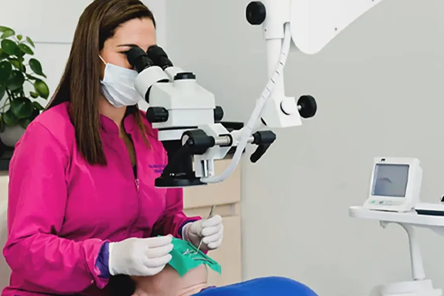
Root canal treatment takes place inside a tooth, in extremely small spaces that may have shapes and anatomical features that are difficult to see with the naked eye. For this reason, the dental operating microscope has become a valuable tool in modern endodontics.
The microscope provides magnification and direct illumination of the working area, allowing the endodontist to visualize small details inside the tooth and work with greater control and precision.
Microscope-assisted root canal treatment is endodontic treatment in which a dental operating microscope is used to magnify and illuminate the inside of the tooth during different stages of the procedure.
A simple way to think about it is as highly specialized magnification combined with focused illumination. The inside of a tooth is very small, and root canals can be narrow, curved, branched, or difficult to locate.
The microscope can reveal details that are difficult to see with the naked eye, making it particularly useful in both initial treatment and more complex cases.
No. Two teeth may look very similar externally while having different internal anatomy. Root canals may be narrow, curved, branched, or located in positions that are difficult to visualize.This is why visualization is so important during root canal treatment.
The dental microscope enlarges the working area and can help the endodontist better understand the internal anatomy of the tooth being treated.
When people hear “microscope,” they often imagine a laboratory. Dental operating microscopes, however, are specifically designed for clinical procedures.
During root canal treatment, the endodontist looks through the microscope while working on the tooth.
The equipment magnifies the image and directs light into the treatment area. This is especially useful because much of endodontic treatment occurs in areas that are not visible to the patient and may be difficult for the dentist to see without magnification.
Modern endodontics uses different tools to support diagnosis and treatment. The dental operating microscope is one of them. Depending on the case, digital radiography, electronic apex locators, diagnostic imaging, and specialized instruments may also be used.
These technologies share a common purpose: to provide information and tools that support careful and precise treatment. Technology does not replace the endodontist’s knowledge, experience, or clinical judgment; it complements them.
Using a microscope does not make root canal treatment more painful. The microscope simply allows the endodontist to view the treatment area with magnification and illumination.
Root canal treatment is performed with local anesthesia to keep the area numb during the procedure.
Temporary sensitivity or discomfort may occur afterward, depending on the patient and the initial condition of the tooth.

When working inside a tooth, small details matter. The dental operating microscope allows us to magnify and illuminate the treatment area and perform procedures with enhanced visualization and precision.
Care available in English, French, and Hebrew